The Morphophysiology of the Cerebral Vascular Event (CVE)
Unidad de Apoyo para el Aprendizaje
Proyecto PAPIME Clave: PE308817
StartLike the rest of our organs, the brain needs oxygen and nutrients provided by the blood, which come through the cerebral arteries, which do not have energy storage systems, so it is necessary that the contribution is constant and permanent.
The cerebrovascular event or CVE occurs when a cerebral artery is obstructed by a blood clot for the supply of oxygen to the brain. Without oxygen, brain tissues die in a few minutes. As a result, the parts of the body that are under the control of these cells stop working properly, causing the appearance of neurological symptoms secondary to the lack of oxygen.
Click to observe some tomographies about the CVE.
In this unit you will be able to recognize in a general way the components that make up the cerebral circulation that, in conjunction with time, are determining factors to locate at what level of the cerebral circulation the damage occurred.
The Cerebral Vascular Event (CVE) has been traditionally defined as a clinical syndrome characterized by the rapid development of symptoms and / or signs usually corresponding to a focal neurological condition, and which persists for more than 24 hours, with no other apparent cause other than vascular origin.

(n. a.) (n. d.). [In: Ischemic Stroke] [graphic]. Retrieved from: https://commons.wikimedia.org/wiki/File:Ischemic_Stroke.svg

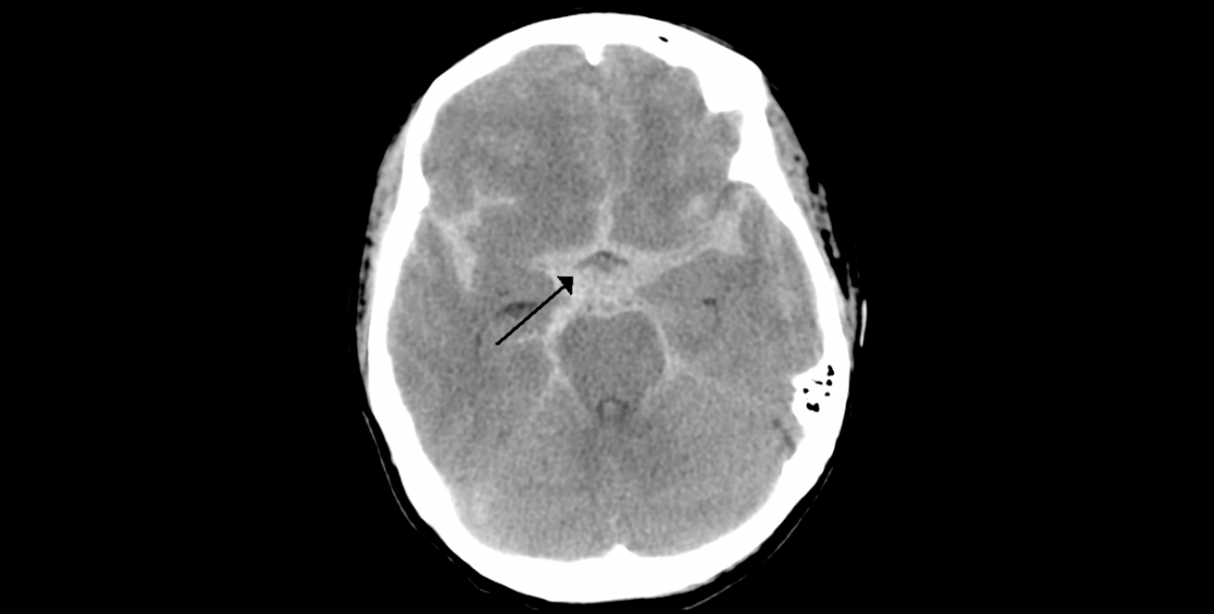
The cerebral ischemia is the consequence of the occlusion of a vessel and may have transient (transient ischemic attack) or permanent manifestations, which implies an irreversible neuronal damage. In intracerebral hemorrhage (ICH), rupture of a vessel results in a blood collection in the cerebral parenchyma or in the subarachnoid space. (Barinagarrementeria and Cantú, 2003)

(n. a.) (n. d.). [In: Brain Ischemia] [Computing axial tomography]. Retrieved from: https://en.wikipedia.org/wiki/Brain_ischemia
Brain ischemia is a dynamic and unstable state where the magnitude of brain tissue damage will depend on three fundamental factors:


Classification
Depending on its duration, ischemia can be transient without producing brain damage when the blood flow is restored promptly, or it can be prolonged until it leads to the destruction of the neurons, leading to cerebral infarction.

Clinical sub-classification
After classifying the ischemia by time, we proceed to perform a much more specific classification, taking into account the place where the damage occurs, either in the carotid portion or in the basilar portion and those that affect the different arteries, speaking at the intracranial and extra cranial level.


Dr. Manuel Porras Bentacourt. (n. d.). Evento vascular cerebral (EVC). Consulted on the 31 of October of 2018 from http://neurochih.com/Inicio/evento-vascular-cerebral-evc/
A hemorrhagic cerebrovascular event (CVE) is a condition that occurs when a blood vessel in the brain breaks down. The blood runs out and can irritate or hurt the brain tissue or cause damage by pushing against neighboring areas. Then you can see a diagram of each one with its corresponding subcategories and characteristics.

Intracerebral hemorrhage occurs when the rupture of a brain vessel results in spontaneous bleeding within the cranial cavity; They are classified according to their location.
Intraparenchymal hemorrhage (HIP) is a blood collection within the brain parenchyma, produced by vascular rupture, whose shape, size, location and etiology is variable. The clinical manifestations of HIP are consistent with the location of the hematoma and depending on the underlying cause of the hemorrhage, the HIP is divided into primary or secondary.
Subarachnoid hemorrhage

Wikipedia. (n.d.). Subarachnoid hemorrhage [Computed axial tomography]. Retrieved from https://en.wikipedia.org/wiki/Subarachnoid_hemorrhage
This type of hemorrhage is related to excessive alcohol consumption, smoking and HBP, in addition to representing an increased risk of SAH for women (Barinagarrementeria and Cantú, 2003).
The importance of knowing the vascularization system is the relation that keeps to diagnose a CVD or some other pathology. It is this system that will provide oxygen and blood to the brain if any obstruction occurs at the level of any structure that composes it. The blood supply would decrease, and the patient's life would play a very important role, where time is a vital factor.
Unlike most organs, whose vascularization system is based on a single vascular pedicle, the brain has a special irrigation composed of four large arteries that form two large well-differentiated vascular systems:

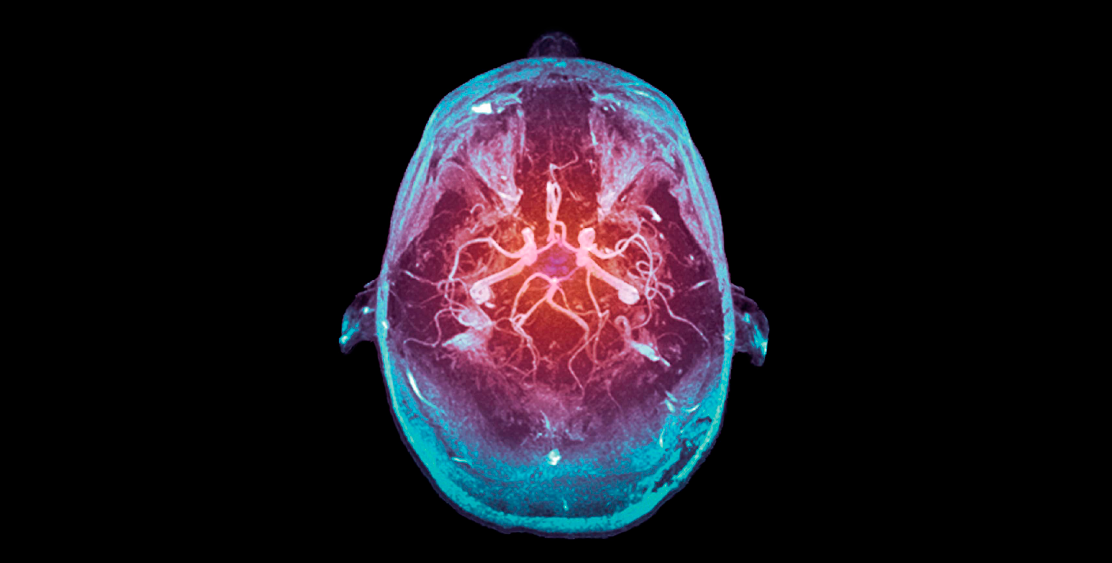
The vertebral arteries fuse intracranially formed a trunk known as the basilar artery. In the following image you can see the emphasis on the regions that irrigate the carotid and vertebrobasilar system:

[Entry: Edema cerebral] [tomography]. Retrieved from: https://commons.wikimedia.org/wiki/File:MRI_brain_tumor.jpg
Click on each region to recognize its structure and description.
The left common carotid is derived from the aortic arch, while the right common carotid is derived from the innominate artery. The most common variations include the anomalous origin of the left common carotid from the innominate or right common carotid of the aortic arch. (Poveda, 2009)
Branches of the common carotids are usually not derived before their bifurcation, although the superior thyroid artery and the ascending pharyngeal artery can be derived from this vessel when the bifurcation occurs at a very high level, usually beyond the body of the third cervical vertebra. (Poveda, 2009)

(Poveda, 2009)
The external carotid artery has eight branches, which usually irrigate extracranial segments. These are: the superior thyroid, the ascending pharyngeal, the lingual, the facial, the occipital, the posterior auricular, the superficial temporal and the maxillary artery. Several of these arteries supply the dura of the basal and lateral brain surface. Said meningeal branches also replace the dura of the posterior fossa in a manner shared with branches of the occipital artery. (Poveda, 2009)

[Entry: External Carotid Artery] [picture]. Retrieved from https://commons.wikimedia.org/wiki/File:Arteria_car%C3%B3tida_externa.png
The vertebral artery is divided into four segments: the initial, the intervertebral, the horizontal, and the intracranial. The branches of the intervertebral segment are the meningeal, the muscular and the radicular. The meninges replace the dura of the foramen magnum; the muscles, the deep muscles of the neck and create anastomosis towards the occipital. The radicular enter the spinal canal and provide collaterals to the anterior and posterior spinal branches of the horizontal segment. This segment also provides small meningeal branches, which replace the dura mater of the posterior fossa and anastomose with its homologous vertebral and branches of the external carotid. The aforementioned spinal branches provide flow to the medulla oblongata (or spinal bulb) and to the posterior spine. The branches of the intracranial segment include the anterior spinal cord and the cerebellar anterior-posterior segment. (Poveda, 2009)
")
The polygon of Willis is formed by the two internal carotids, the two segments of the anterior cerebral artery, the anterior, posterior communicating arteries and the posterior cerebral arteries. It usually provides communication between the posterior and anterior circulation, although this normal conformation is observed in 50% of patients. The most common variations are hypoplasia of the left posterior communicator, branches of the posterior communicating, parallel to the anterior choroidal, the left posterior communicating originating from the left internal carotid artery and the absence of the right posterior communicating artery.

Wikipedia. [Entry: Cerebral Artery Circuit] [picture]. Retrieved from: https:en.wikipedia.org/wiki/Circle_of_Willis
*With a Doctor's Letter. (April 18, 2017). A BLOOD VASCULARIZATION PART 1: carotid internal [Video file]. Consulted on https://www.youtube.com/watch?v=P9tumdhT0Y4&t=109s&list=LLQDMz-7G-3u hAYhlTx3gig&index=1
*With a Doctor's Letter. (May 1, 2017). BLOOD ARTERIAL VASCULARIZATION PART 2: vertebrobasilar [Video file]. Consulted on https://www.youtube.com/watch?v=7f6VjMIsYIg

Risk factors
Click on each risk factor to review its impact and relationship with the CVE.

According to the occluded vessel, the clinical presentation of the patients varies, and this can be framed within different clinical syndromes.
There are different signs and symptoms that appear in the EVC, which will trigger the appearance of different clinical manifestations. The main symptoms and signs of cerebral ischemia in the carotid and vertebrobasilar territory are summarized in the following diagrams.
Click on each clinical sign and symptom to recognize its brain structure.

It is essential to recognize the composition of the clinical syndrome called "cerebrovascular event" (EVC) in order to proceed correctly and make the decisions that correspond to the situation that arises, because without the oxygen supply the cerebral tissues die in a few minutes.
The fatality in the presentation of the cerebral vascular event depends on several factors, among which the age and the previous health condition stand out.

The CVE is a neurological disorder that is characterized by its sudden onset, usually without warning, with symptoms of 24 hours or more, causing severe sequelae or even death.
Information sources
Bibliography
Barinagarrementeria, F. & Cantú, G. (2003). Enfermedad vascular cerebral (3.ª ed.) (pp. 1-10 y 23-44). México: El Manual Moderno.
Sancho, J. & Rodriguez, A. (2012). Manual del residente de neurología (pp. 1307 y 1647-1667). Madrid: ENE Life Publicidad S. A.
Electronic documents
Arana, A., Uribe, C. S., Muñoz, A., Salinas F. A. & Celis, J. I. (n. d.). Enfermedad cerebrovascular [Versión electrónica]. Consulted on the 31 of October of 2018 from http://www.medynet.com/usuarios/jraguilar/Enfermedad%20cerebrovascular.pdf
Arauz, A. & Ruíz-Franco, A. (2012, May-June). Enfermedad vascular cerebral (3.ª ed.) [Versión electrónica]. Revista de la Facultad de Medicina de la UNAM, 55(3), pp. 11-21. Consulted on the 31 of October of 2018 from http://www.scielo.org.mx/pdf/facmed/v55n3/v55n3a3.pdf
Cazares, K. (2015, July-September). La enfermedad vascular cerebral en México: un problema de salud en incremento [Versión electrónica]. Anales de Radiología México, 14(3). Consulted on the 11 of January of 2018 from: http://www.medigraphic.com/pdfs/anaradmex/arm-2015/arm153a.pdf
Poveda, J. (2009, July-December). Anatomía básica cerebral para el cardiólogo intervencionista [Versión electrónica]. Revista costarricense de cardiología, 11(2), pp. 33-40. Consulted on the 31 of October of 2018 from http://www.scielo.sa.cr/pdf/rcc/v11n2/a10v11n2.pdf
How to quote
Flores, C. & Rodríguez, G. (2019). The morphophysiology of the cerebral vascular event (CVE). Unidades de Apoyo para el Aprendizaje. CUAED/ENEO-UNAM. Retrieved on (date) from (link)